
The integration of machine learning and artificial intelligence (AI) is providing a new lens for viewing retinal health, transforming the field of ophthalmology from a reactive one to proactive and predictive. Retinal scans offer a non-invasive view of blood vessels and nerve fibres. They are not just a window into the eye, but also a valuable diagnostic tool for a host of diseases.
For instance, the narrowing of small blood vessels in the retina called retinal arterioles is related to long-term risk of high blood pressure, while larger diameter or width of retinal veins is related to kidney issues in people with Type-1 diabetes. Moreover, arteriolar-to-venular diameter ratio is an established biomarker for stroke and heart diseases.
The retina thus provides a unique opportunity to assess and diagnose various ailments such as diabetes mellitus, coronary heart disease, high blood pressure, kidney disease, and neurodegenerative disorders. This is because the structure of retinal vessels can be considered a witness of the patient’s vascular status. With an increase in the aging population and poor lifestyle choices, the prevalence of these diseases is on the rise. Early diagnosis and identifying high-risk individuals is the need of the hour.
READ I India must emulate Japan’s compact city model
AI retinal scans: A non-invasive health mirror
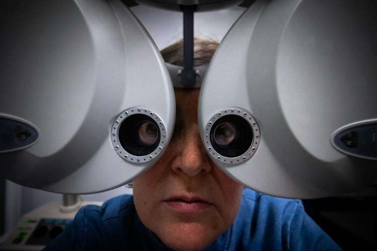
The last two decades have witnessed a growing interest in imaging of the blood vessels of the retina. Technology to capture retinal images such as retinal fundus photography, optical coherence tomography–angiography (OCT-A) or adaptive optics have made it possible to get accurate data on our circulatory system.
Fundus photography is used to capture images of the inside of the eye which includes structures such as the retina, optic nerve head, macula, retinal blood vessels, choroid and the vitreous.
These images are used to screen and detect various causes of treatable and preventable blindness such as diabetic retinopathy, age-related macular degeneration, and glaucoma, among others.
OCT-A is used to obtain detailed visuals of the vascular networks of the retina; it is non-invasive, time-efficient, and allows for a three-dimensional examination of the retina.
Over the last decade, much research has been going on to develop software that can enable an automatic analysis of the retinal vascular network from these imaging techniques to provide an accurate description of the patient’s arteries and veins.
Recently, a new approach called “oculomics”, that uses retinal image datasets and artificial intelligence algorithms, has increased interest in retinal microvascular biomarkers.
Generative AI and eye surgery
A common problem that AI can help resolve in the field of ophthalmology is improving surgical outcomes for patients with macular holes, a condition that causes central vision loss.
Macular holes are defects in the macula, a part of the retina. Those who have the disease have problems seeing clearly, especially in their central field of vision.
Surgery to treat a macular hole, called vitrectomy, has high success rates if the hole is small.
Despite being the standard treatment for the disease, the success of the surgery can vary — a failed macular hole surgery often requires another attempt, increased expenditure and emotional stress for the patient.
Here, AI tools that can learn from pre- and post-operative images can be leveraged. The technology can help predict what a patient’s retina will look like after surgery, including the likelihood of the macular hole closing.
This predictive capability is a significant leap forward, providing a powerful tool for surgeons to plan the procedure appropriately and counsel patients before surgery, helping them make more informed decisions and setting accurate expectations.
Non-invasive screening for diabetes
A second, equally impactful project being worked on by this author and her team is motivated by the need for more accessible and non-invasive diagnostic tools for diabetes.
Current screening methods for glycated haemoglobin (HbA1c) levels — the test measures average blood sugar levels over the past 90 days, expressed as a percentage — typically require blood samples, which can be inconvenient and create barriers to care.
This is a particularly critical issue for India, which is now considered the diabetes capital of the world.
As per the International Diabetes Federation (IDF) Atlas 11th edition, India surpassed China with the highest number of diabetic individuals globally, and the number is predicted to increase by 75 percent in the next 25 years.
This highlights the urgent need for a scalable, cost-effective solution that obviates the need for a blood test. Researchers on this project are developing a deep learning framework that can classify HbA1c levels directly from retinal images.
The designed model is highly robust and accurate — the program has learned to identify patterns in eye images that are connected to a person’s average blood sugar level (HbA1c).
Depending on the patterns, it can give a simple “Yes/No” answer on whether blood sugar is in a healthy range. It can also provide a more detailed report that classifies the levels as optimal, elevated, or high risk.
The technology can be deployed as a user-friendly application, which can be used for mass screening, making it more cost-effective than traditional blood tests for the country’s large diabetic population.
This innovative approach could transform routine diabetes screening, allowing for earlier detection and intervention, without the need for traditional blood tests.
Unified framework for disease classification
Many systemic conditions such as high blood sugar and cholesterol manifest in the retina with subtle signs that appear before other clinical symptoms.
This author and her team are working to tackle the broader challenge of classifying multiple diseases from a retinal image.
The project utilises Auxiliary Classifier Generative Adversarial Networks (AC-GANs), which are particularly effective for disease classification.
The AC-GAN framework not only generates realistic retinal images to augment limited datasets but also trains a classifier to differentiate between eye diseases, and systemic diseases such as those involving the heart and the kidney.
This dual-purpose system has the potential to streamline diagnostics, allowing clinicians to screen for a wide array of conditions through a single, efficient imaging session.
Together, these projects represent a new era of AI-driven ophthalmology, where retinal scans become a comprehensive health report, offering unprecedented insights into both eye and body health.
Several researchers globally are using AI to check for eye diseases, but applications such as predicting a person’s average blood sugar level from an eye scan, or the development of a single tool that can screen for multiple conditions – in the eye, and throughout the body — is not only unique, but also crucial, especially for low-resources countries such as India.
Black box and other hurdles
Despite the exciting potential, there are hurdles to overcome. One of the major challenges is to get enough patient data from different backgrounds to make sure the AI is accurate.
There’s also the “black box” problem — AI’s decision-making process is hard for doctors to understand, which can make them hesitant to trust it.
The challenges are being tackled — researchers are sharing anonymous data across different hospitals to create larger, more diverse datasets. They are also working on ways to make AI more transparent by showing what specific parts of the eye it is looking at to make the diagnosis. These efforts are helping to build trust and ensure the AI tools are both safe and effective for real-world use.
Dr Devanjali Relan is Professor, BML Munjal University. This research is being conducted through a collaboration between Dr Relan and Dr Dhanashree Ratra and her team at Sankara Nethralaya, Chennai, India. Originally published under Creative Commons by 360info .
.